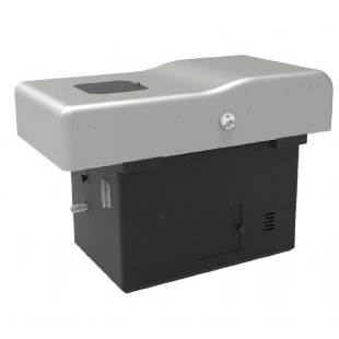
大组织光片扫描显微镜
产品名称: 大组织光片扫描显微镜
英文名称: Large tissue light slice scanning microscopy
产品编号: LSI XL
产品价格: 0
产品产地: 新加坡
品牌商标: LogiSci
更新时间: 2024-05-07T09:25:55
使用范围: null
<p>&lt;p&gt;&amp;lt;p class="ql-align-justify"&amp;gt;LSI XL系列光片扫描显微镜旨在以高分辨率高速对大型样品进行三维成像。该系统利用线性贝塞尔光片技术,配合LSI独有的四面照明技术,可提供市场上最均匀的样品照明。LSI XL系列光片扫描显微镜配备了折射率(RI)校正光学器件,可在1.33至1.56之间调节,以确保在各种浸没介质中的最佳成像质量。可更换的样品室可容纳2cmx2cm的样品。&amp;lt;/p&amp;gt;&amp;lt;p class="ql-align-justify"&amp;gt;&amp;lt;br&amp;gt;&amp;lt;/p&amp;gt;&amp;lt;p class="ql-align-justify"&amp;gt;LSI XL系列光片扫描显微镜的应用包括对通过日前广泛应用的以水基或溶剂基方法处理的大型透明组织或器官样品进行成像,以及通过内置的一键化多位点成像功能对大量活体透明样品(如斑马鱼或果蝇胚胎)的拍摄。&amp;lt;/p&amp;gt;&amp;lt;p class="ql-align-justify"&amp;gt;&amp;lt;br&amp;gt;&amp;lt;/p&amp;gt;&amp;lt;p class="ql-align-justify"&amp;gt; &amp;lt;/p&amp;gt;&amp;lt;p class="ql-align-justify"&amp;gt;&amp;lt;br&amp;gt;&amp;lt;/p&amp;gt;&amp;lt;p class="ql-align-center"&amp;gt;&amp;lt;img src="https://nwzimg.wezhan.cn/contents/sitefiles2053/10267688https://msimg.bioon.com/bionline//images/28598716.png"&amp;gt;&amp;lt;/p&amp;gt;&amp;lt;p class="ql-align-center"&amp;gt; &amp;lt;/p&amp;gt;&amp;lt;p class="ql-align-center"&amp;gt;&amp;lt;br&amp;gt;&amp;lt;/p&amp;gt;&amp;lt;p class="ql-align-justify"&amp;gt;&amp;lt;strong&amp;gt;主要特点&amp;lt;/strong&amp;gt;&amp;lt;/p&amp;gt;&amp;lt;p class="ql-align-justify"&amp;gt;&amp;lt;br&amp;gt;&amp;lt;/p&amp;gt;&amp;lt;p class="ql-align-justify"&amp;gt;*线性贝塞尔光片和RI矫正光学模组提供了最佳的成像效果,分辨率可达500nm。&amp;lt;/p&amp;gt;&amp;lt;p class="ql-align-justify"&amp;gt;&amp;lt;br&amp;gt;&amp;lt;/p&amp;gt;&amp;lt;p class="ql-align-justify"&amp;gt;*独特的四侧照明技术可显着增加照明深度和均匀度,尤其适合对在大型透明化样品成像。&amp;lt;/p&amp;gt;&amp;lt;p class="ql-align-justify"&amp;gt;&amp;lt;br&amp;gt;&amp;lt;/p&amp;gt;&amp;lt;p class="ql-align-justify"&amp;gt;*适用于活体胚胎的长时间成像,专为大型透明化样品设计的光片成像平台,同时也可完美得适用于活体斑马鱼或果蝇胚胎的成像,其通量比传统的光片显微镜高得多。&amp;lt;/p&amp;gt;&amp;lt;p class="ql-align-justify"&amp;gt;&amp;lt;br&amp;gt;&amp;lt;/p&amp;gt;&amp;lt;p class="ql-align-justify"&amp;gt;*智能化易用的软件系统配有快速数据处理功能,同时内置了3D渲染,多位置采集及自动拼接和反卷积等图像分析功能。&amp;lt;/p&amp;gt;&amp;lt;p class="ql-align-justify"&amp;gt;&amp;lt;br&amp;gt;&amp;lt;/p&amp;gt;&amp;lt;p class="ql-align-justify"&amp;gt;*一体化台面紧凑设计配有内置隔振系统,无需外置隔振台。&amp;lt;/p&amp;gt;&amp;lt;p class="ql-align-justify"&amp;gt;&amp;lt;br&amp;gt;&amp;lt;/p&amp;gt;&amp;lt;p class="ql-align-justify"&amp;gt; &amp;lt;/p&amp;gt;&amp;lt;p class="ql-align-justify"&amp;gt;&amp;lt;br&amp;gt;&amp;lt;/p&amp;gt;&amp;lt;p class="ql-align-center"&amp;gt;&amp;lt;img src="https://nwzimg.wezhan.cn/contents/sitefiles2053/10267688https://msimg.bioon.com/bionline//images/28598715.png"&amp;gt;&amp;lt;/p&amp;gt;&amp;lt;p class="ql-align-center"&amp;gt; &amp;lt;/p&amp;gt;&amp;lt;p class="ql-align-center"&amp;gt;&amp;lt;br&amp;gt;&amp;lt;/p&amp;gt;&amp;lt;p class="ql-align-justify"&amp;gt;&amp;lt;strong&amp;gt;线性贝塞尔光片(LSI)技术&amp;lt;/strong&amp;gt;&amp;lt;/p&amp;gt;&amp;lt;p class="ql-align-justify"&amp;gt;&amp;lt;br&amp;gt;&amp;lt;/p&amp;gt;&amp;lt;p class="ql-align-justify"&amp;gt;LSI技术通过物理和光学调制获取的光片,远比传统的高斯光片薄,有效长度也更长。因此LSI显微镜不仅具有极低的光毒率和超快的成像速度的特点,而且其出色的三维分辨率和高信噪比令其具有机器出色的层切能力&amp;lt;/p&amp;gt;&amp;lt;p class="ql-align-justify"&amp;gt;&amp;lt;br&amp;gt;&amp;lt;/p&amp;gt;&amp;lt;p class="ql-align-justify"&amp;gt; &amp;lt;/p&amp;gt;&amp;lt;p class="ql-align-justify"&amp;gt;&amp;lt;br&amp;gt;&amp;lt;/p&amp;gt;&amp;lt;p class="ql-align-center"&amp;gt;&amp;lt;img src="https://nwzimg.wezhan.cn/contents/sitefiles2053/10267688https://msimg.bioon.com/bionline//images/28599233.jpg"&amp;gt;&amp;lt;/p&amp;gt;&amp;lt;p class="ql-align-center"&amp;gt; &amp;lt;/p&amp;gt;&amp;lt;p class="ql-align-center"&amp;gt;&amp;lt;br&amp;gt;&amp;lt;/p&amp;gt;&amp;lt;p class="ql-align-justify"&amp;gt;&amp;lt;strong&amp;gt;应用领域&amp;lt;/strong&amp;gt;&amp;lt;/p&amp;gt;&amp;lt;p class="ql-align-justify"&amp;gt;&amp;lt;br&amp;gt;&amp;lt;/p&amp;gt;&amp;lt;p class="ql-align-justify"&amp;gt;&amp;lt;strong style="color: rgb(0, 0, 0);"&amp;gt;神经示踪三维成像&amp;lt;/strong&amp;gt;&amp;lt;/p&amp;gt;&amp;lt;p class="ql-align-justify"&amp;gt;&amp;lt;br&amp;gt;&amp;lt;/p&amp;gt;&amp;lt;p class="ql-align-justify"&amp;gt; &amp;lt;/p&amp;gt;&amp;lt;p class="ql-align-justify"&amp;gt;&amp;lt;br&amp;gt;&amp;lt;/p&amp;gt;&amp;lt;p class="ql-align-center"&amp;gt;&amp;lt;strong style="color: rgb(0, 0, 0);"&amp;gt;&amp;lt;img src="https://nwzimg.wezhan.cn/contents/sitefiles2053/10267688https://msimg.bioon.com/bionline//images/28410790.png"&amp;gt;&amp;lt;/strong&amp;gt;&amp;lt;/p&amp;gt;&amp;lt;p class="ql-align-center"&amp;gt; &amp;lt;/p&amp;gt;&amp;lt;p class="ql-align-center"&amp;gt;&amp;lt;br&amp;gt;&amp;lt;/p&amp;gt;&amp;lt;p&amp;gt;&amp;lt;strong style="color: rgb(0, 0, 0);"&amp;gt;全脑神经胞体三维成像&amp;lt;/strong&amp;gt;&amp;lt;/p&amp;gt;&amp;lt;p&amp;gt;&amp;lt;br&amp;gt;&amp;lt;/p&amp;gt;&amp;lt;p&amp;gt; &amp;lt;/p&amp;gt;&amp;lt;p&amp;gt;&amp;lt;br&amp;gt;&amp;lt;/p&amp;gt;&amp;lt;p class="ql-align-center"&amp;gt;&amp;lt;strong style="color: rgb(0, 0, 0);"&amp;gt;&amp;lt;img src="https://nwzimg.wezhan.cn/contents/sitefiles2053/10267688https://msimg.bioon.com/bionline//images/28495932.png"&amp;gt;&amp;lt;/strong&amp;gt;&amp;lt;/p&amp;gt;&amp;lt;p class="ql-align-center"&amp;gt; &amp;lt;/p&amp;gt;&amp;lt;p class="ql-align-center"&amp;gt;&amp;lt;br&amp;gt;&amp;lt;/p&amp;gt;&amp;lt;p&amp;gt;&amp;lt;strong style="color: rgb(0, 0, 0);"&amp;gt;全脑血管三维成像&amp;lt;/strong&amp;gt;&amp;lt;/p&amp;gt;&amp;lt;p&amp;gt;&amp;lt;br&amp;gt;&amp;lt;/p&amp;gt;&amp;lt;p&amp;gt; &amp;lt;/p&amp;gt;&amp;lt;p&amp;gt;&amp;lt;br&amp;gt;&amp;lt;/p&amp;gt;&amp;lt;p class="ql-align-center"&amp;gt;&amp;lt;strong style="color: rgb(0, 0, 0);"&amp;gt;&amp;lt;img src="https://nwzimg.wezhan.cn/contents/sitefiles2053/10267688https://msimg.bioon.com/bionline//images/28410798.png"&amp;gt;&amp;lt;/strong&amp;gt;&amp;lt;/p&amp;gt;&amp;lt;p class="ql-align-center"&amp;gt; &amp;lt;/p&amp;gt;&amp;lt;p class="ql-align-center"&amp;gt;&amp;lt;br&amp;gt;&amp;lt;/p&amp;gt;&amp;lt;p&amp;gt;&amp;lt;strong style="color: rgb(0, 0, 0);"&amp;gt;动物胚胎成像&amp;lt;/strong&amp;gt;&amp;lt;/p&amp;gt;&amp;lt;p&amp;gt;&amp;lt;br&amp;gt;&amp;lt;/p&amp;gt;&amp;lt;p class="ql-align-justify"&amp;gt;&amp;lt;span style="color: rgb(0, 0, 0);"&amp;gt;同时可进行亚细胞分辨率的完整哺乳动物大脑中枢和外周神经系统的发育及微循环三维介观形态学图谱等研究。&amp;lt;/span&amp;gt;&amp;lt;/p&amp;gt;&amp;lt;p class="ql-align-justify"&amp;gt;&amp;lt;br&amp;gt;&amp;lt;/p&amp;gt;&amp;lt;p class="ql-align-justify"&amp;gt; &amp;lt;/p&amp;gt;&amp;lt;p class="ql-align-justify"&amp;gt;&amp;lt;br&amp;gt;&amp;lt;/p&amp;gt;&amp;lt;p class="ql-align-center"&amp;gt;&amp;lt;span style="color: rgb(0, 0, 0);"&amp;gt;&amp;lt;img src="https://nwzimg.wezhan.cn/contents/sitefiles2053/10267688https://msimg.bioon.com/bionline//images/28599658.jpg"&amp;gt;&amp;lt;/span&amp;gt;&amp;lt;/p&amp;gt;&amp;lt;p class="ql-align-center"&amp;gt; &amp;lt;/p&amp;gt;&amp;lt;p class="ql-align-center"&amp;gt;&amp;lt;br&amp;gt;&amp;lt;/p&amp;gt;&amp;lt;p&amp;gt;&amp;lt;strong style="color: rgb(0, 0, 0);"&amp;gt;微循环血管三维成像&amp;lt;/strong&amp;gt;&amp;lt;/p&amp;gt;&amp;lt;p&amp;gt;&amp;lt;br&amp;gt;&amp;lt;/p&amp;gt;&amp;lt;p&amp;gt; &amp;lt;/p&amp;gt;&amp;lt;p&amp;gt;&amp;lt;br&amp;gt;&amp;lt;/p&amp;gt;&amp;lt;p class="ql-align-center"&amp;gt;&amp;lt;strong style="color: rgb(0, 0, 0);"&amp;gt;&amp;lt;img src="https://nwzimg.wezhan.cn/contents/sitefiles2053/10267688https://msimg.bioon.com/bionline//images/28599856.png"&amp;gt;&amp;lt;/strong&amp;gt;&amp;lt;/p&amp;gt;&amp;lt;p class="ql-align-center"&amp;gt; &amp;lt;/p&amp;gt;&amp;lt;p class="ql-align-center"&amp;gt;&amp;lt;br&amp;gt;&amp;lt;/p&amp;gt;&amp;lt;p&amp;gt;&amp;lt;strong style="color: rgb(0, 0, 0);"&amp;gt;三维肿瘤病理成像&amp;lt;/strong&amp;gt;&amp;lt;/p&amp;gt;&amp;lt;p&amp;gt;&amp;lt;br&amp;gt;&amp;lt;/p&amp;gt;&amp;lt;p&amp;gt; &amp;lt;/p&amp;gt;&amp;lt;p&amp;gt;&amp;lt;br&amp;gt;&amp;lt;/p&amp;gt;&amp;lt;p class="ql-align-center"&amp;gt;&amp;lt;strong style="color: rgb(0, 0, 0);"&amp;gt;&amp;lt;img src="https://nwzimg.wezhan.cn/contents/sitefiles2053/10267688https://msimg.bioon.com/bionline//images/28599857.png"&amp;gt;&amp;lt;/strong&amp;gt;&amp;lt;/p&amp;gt;&amp;lt;p class="ql-align-center"&amp;gt; &amp;lt;/p&amp;gt;&amp;lt;p class="ql-align-center"&amp;gt;&amp;lt;br&amp;gt;&amp;lt;/p&amp;gt;&amp;lt;p&amp;gt;&amp;lt;strong style="color: rgb(0, 0, 0);"&amp;gt;三维肿瘤病理应用实例&amp;lt;/strong&amp;gt;&amp;lt;/p&amp;gt;&amp;lt;p&amp;gt;&amp;lt;br&amp;gt;&amp;lt;/p&amp;gt;&amp;lt;p&amp;gt; &amp;lt;/p&amp;gt;&amp;lt;p&amp;gt;&amp;lt;br&amp;gt;&amp;lt;/p&amp;gt;&amp;lt;p&amp;gt;&amp;lt;strong style="color: rgb(0, 0, 0);"&amp;gt;&amp;lt;img src="https://nwzimg.wezhan.cn/contents/sitefiles2053/10267688https://msimg.bioon.com/bionline//images/28599858.png"&amp;gt;&amp;lt;/strong&amp;gt;&amp;lt;/p&amp;gt;&amp;lt;p&amp;gt;&amp;lt;br&amp;gt;&amp;lt;/p&amp;gt;&amp;lt;p&amp;gt;&amp;lt;br&amp;gt;&amp;lt;/p&amp;gt;&amp;lt;p&amp;gt; &amp;lt;/p&amp;gt;&lt;/p&gt;</p>