尼康 Nikon A1 MP+/A1R MP+双光子显微镜
产品名称: 尼康 Nikon A1 MP+/A1R MP+双光子显微镜
英文名称:
产品编号: A1MP+/A1RMP+
产品价格: 0
产品产地: 日本
品牌商标: Nikon/尼康
更新时间: null
使用范围: null
Amazingly deep — A1 MP+/A1R MP+ sharply visualize ultra-deep dynamics within living organisms.
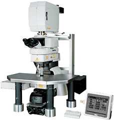
 Configured with Ni-E
The A1 MP+/A1R MP+ multiphoton confocal microscopes provide faster and sharper imaging from deeper within living organisms, extending the boundaries of traditional research techniques in biological sciences. They are compatible with both upright and inverted microscopes, and provide optimum multiphoton imaging configurations for brain research, other neuroscience applications and in vivo imaging of living specimens.
Ultrahigh-speed imaging of up to 420 frames per second
The Nikon resonant scanner is equipped with A1R MP+ and capable of high-speed 420 fps (512 x 32 pixels) imaging, the world's fastest for a multiphoton microscope using point scanning technology. Unique to this design is a resonant scan mirror capability that enables imaging full fields of view at much higher speeds than with conventional galvano scanners. Nikon's optical pixel clock system, which monitors the position of the resonant mirror in real time, adjusts the pixel clock to ensure more stable, geometrically correct and more evenly illuminated imaging, even at high speeds. This enables the successful visualization of in vivo rapid changes, such as reactions in living organisms, dynamics and cell interactions.
- Visualization of intravital microcirculation
Blood cells in blood vessels within a living organism were excited by a femtosecond pulsed IR laser with the A1R MP+' ultrahigh-speed resonant scanner, and their movements were simultaneously captured in three successive fluorescence images at 30 fps (30 msec), with three separate color channels.
Three fluorescent probes are simultaneously excited and imaged–nucleus (blue), endothelium (green), and plasma (red). The long-wavelength ultrafast laser in combination with the ultrahigh-speed resonant scanner effectively reduces photodamage and makes time resolved multiphoton imaging of biomolecules possible. - Image resolution: 512 x 512 pixels, Image acquisition speed: 30 fps, Objective: water immersion objective 60x
- Imaged with the cooperation of: Dr. Satoshi Nishimura, Department of Cardiovascular Medicine, the University of Tokyo, TSBMI, the University of Tokyo, PRESTO, Japan Science and Technology Agency
Deep specimen imaging with non-descanned detectors (NDD)
The fluorescence emissions from deep within a specimen are highly scattered in multiphoton excitation, and therefore the conventional detector using a pinhole cannot provide bright fluorescent images. The episcopic NDD in the A1 MP+/A1R MP+ is located close to the back aperture of the objective to detect the maximum amount of scattered emission signals from deep within living specimens. The use of this four-channel detector in combination with special spectral mirrors, together with Nikon's unmixing algorithm, eliminates cross talk between fluorescent probes with highly overlapping emission spectra. Background auto-fluorescence is also eliminated, enabling high-contrast image capture from deep within the specimen. Using diascopic NDD* together with episcopic NDD, brighter images can be acquired by detecting fluorescence signals from both reflected and transmitted.
- *Compatible with Ni-E focusing nosepiece microscope

4-channel episcopic NDD

4-channel diascopic NDD

- In vivo image of deep areas of cerebral cortex of a mouse
The cerebral cortex of an H-line 5-week-old mouse was studied with the open skull method. The entire shape of dendrites of pyramidal cells in layer V expressing EYFP were visualized from the bottom layer into a superficial layer. In addition, the fluorescence signal of white matter in deeper areas was also studied. - Left) 3D reconstruction image
Right) Z-stack images- Top:
- dendrites located in superficial layers in the layer V pyramidal cells
25 µm from the surface - Middle:
- basal dendrites in the layer V pyramidal cells
625 µm from the surface - Bottom:
- fluorescence from white matter
- Excitation wavelength: 930 nm
Objective: CFI75 Apo 25xW MP (NA 1.10 WD 2.0)
Excitation wavelength: 930 nm
Objective: CFI75 Apo 25xW MP (NA 1.10 WD 2.0) - Photographed with the cooperation of:
Dr. Tomomi Nemoto, Research Institute for Electronic Science, Hokkaido University
Dr. Shigenori Nonaka, National Institute for Basic Biology
Dr. Takeshi Imamura, Graduate School of Medicine, Ehime University


- Mouse cerebral cortex multi-color imaging
Simultaneous acquisition of three channels in anesthetized YFP-H mouse using IR excitation of 950 nm and imaging Second Harmonic Generation (SHG) and two fluorescence emissions.
Cyan: SHG signal of dura mater
Yellow: EYFP pyramidal neurons in layer V of the cortex
Red: SRB-labeled blood vessels - Photographed with the cooperation of:
Dr. Ryosuke Kawakami, Dr. Terumasa Hibi, Dr. Tomomi Nemoto, Research Institute for Electronic Science, Hokkaido University
Super high-sensitive GaAsP NDD ![[New]](/ncgs_common/http://www.nikon.com/products/instruments/lineup/bioscience/confocal/multiphoton/a1rmp/img/icon/new.png)

The newly developed gallium arsenide phosphide (GaAsP) NDD* has approximately twice the sensitivity of a standard NDD and allows clear imaging of deeper areas of in vivo mouse brain over 1.2mm. Its ability to acquire bright images enables faster imaging and higher quality Z-stack imaging. Its high sensitivity allows acquisition of fluorescent signals with less laser power, resulting in less photo damage to living specimens.
- *Compatible with FN1 fixed stage microscope
- Deep brain imaging in in vivo mouse
In vivo imaging of an anesthetized YFP-H mouse (4-week-old) via open skull method. Visualization of the entire layer V pyramidal neurons and the deeper hippocampal neurons. Deep imaging achieved for 3-dimensional imaging of hippocampal dendrites over 1.1 mm into the brain. - Captured with episcopic GaAsP NDD and CFI75 Apochromat 25xW MP objective lens (NA 1.10, WD 2.0 mm)
Photographed with the cooperation of:
Dr. Ryosuke Kawakami, Dr. Terumasa Hibi, Dr. Tomomi Nemoto, Research Institute for Electronic Science, Hokkaido University
Nikon's high-NA objectives are ideal for multiphoton imaging
High-NA objectives have been developed that highly correct chromatic aberrations over a wide wavelength range, from ultraviolet to infrared. Transmission is increased through the use of Nikon's exclusive Nano Crystal Coat technology. In particular, the CFI Apochromat 25xW MP objective lens provides an industry leading highest numerical aperture of 1.10 while still maintaining a 2.0 mm working distance. It also has a collar that corrects chromatic aberrations depending on the depth of the specimen and a 33° manipulator pipette access angle, making it ideal for deep multiphoton imaging and physiology research applications.
- Nano Crystal Coat is a Nikon exclusive lens coating technology using an ultralow refractive index nanoparticle thin film originally developed for the semiconductor fabrications industry. The Nano Crystal Coat particle structure dramatically reduces stray reflections and boosts transmission over a wide wavelength range, producing images with higher signal-to-noise (S/N) ratios.

CFI75 Apochromat 25xW MP
CFI Apochromat LWD 40xWI λS

CFI Apochromat 40xWI λS
CFI Plan Apochromat IR 60xWI
| CFI75 Apochromat 25xW MP | NA 1.1 WD 2.0 Nano Crystal Coat |
|---|---|
| CFI Apochromat LWD 40xWI λS | NA 1.15 WD 0.6 Nano Crystal Coat |
| CFI Apochromat 40xWI λS | NA 1.25 WD 0.18 Nano Crystal Coat |
| CFI Plan Apochromat IR 60xWI | NA 1.27 WD 0.17 Nano Crystal Coat |
Auto laser alignment when changing multiphoton excitation wavelength

When the multiphoton laser wavelength or group velocity dispersion pre-compensation is changed, the multiphoton laser beam positional pointing at the objective back aperture may also change, resulting in uneven intensity across the image, or a slight misalignment between the IR and visible laser light paths.
Verifying the IR laser beam pointing and setting the alignment has traditionally been difficult. Nikon's A1 MP+ series' auto laser alignment function, housed in the Incident Optical Unit for the multiphoton excitation light path, automatically maximizes IR laser alignments with a single click in NIS-Elements C.
Two types of scanning head enable high-speed, high-quality imaging
 A1R MP+ hybrid scanner
A1 MP+ equipped with a galvano (non-resonant) scanner enables high-resolution imaging of up to 4096 x 4096 pixels and high-speed acquisition of 10 fps (512 x 512 pixels). A1R MP+ is a hybrid scanning head that incorporates both a galvano scanner and an ultrahigh-speed resonant scanner. A1R MP+ allows imaging and photoactivation at ultrafast speeds at 420 fps (512 x 32 pixels) necessary for revealing cell dynamics and interaction.
NIS-Elements C acquisition and analysis software
- Simple operations common with Nikon confocal microscopes.


Three color simultaneous fluorescent imaging with 850 nm pulsed IR excitation
(left: before unmixing, right: after unmixing)
- A1 MP+/A1R MP+ and NIS Elements C support triggering applications. This is effective for synchronizing frame and scanning times with electrophysiology recordings, or to externally trigger the confocal to scan.